Bassett Collection of Stereoscopic Images of Human Anatomy
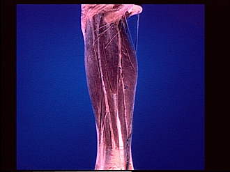
Dissection of lateral aspect of left leg
Muscles of leg, lateral view
Image #193-1
KEYWORDS: Fascia, Leg, Muscles and tendons.
Creative Commons
Stanford holds the copyright to the David L. Bassett anatomical images and has assigned Creative Commons license Attribution-Share Alike 4.0 International to all of the images.

For additional information regarding use and permissions, please contact the Medical History Center.


Dissection of lateral aspect of left leg
Muscles of leg, lateral view
The tela subcutanea has been cut away and the crural fascia (18) has been removed above the region of the ankle. However, the cutaneous branches of the peroneal nerve (6,9,10) have been preserved distal to their points of exit through the crural fascia. These branches have been placed across the underlying muscles in positions that approximately parallel their original peripheral courses.
- Biceps femoris muscle (insertion)
- Tibial tuberosity
- Extensor digitorum longus muscle
- Tibialis anterior muscle
- Anterior intermuscular septum
- Superficial peroneal nerve (dorsal medial cutaneous nerve)
- Peroneus tertius muscle
- Tendon of extensor hallucis longus muscle (visible deep to crural fascia)
- Superficial peroneal nerve (dorsal intermediate cutaneous nerve)
- Lateral sural cutaneous nerve
- Common peroneal branch
- Lesser saphenous vein
- Gastrocnemius muscle
- Soleus muscle
- Posterior intermuscular septum
- Peroneus longus muscle
- Peroneus brevis muscle
- Crural fascia (blending below with superior extensor retinaculum)
- Tendo calcaneus (Achilles)