
Bassett Collection of Stereoscopic Images of Human Anatomy
Dissection of anterior aspect of left leg
Muscles of left leg and foot, anterior view
Image #191-7
KEYWORDS: Foot and toes, Leg, Muscles and tendons.
Creative Commons
Stanford holds the copyright to the David L. Bassett anatomical images and has assigned Creative Commons license Attribution-Share Alike 4.0 International to all of the images.

For additional information regarding use and permissions, please contact the Medical History Center.
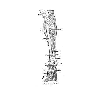
Dissection of anterior aspect of left leg
Muscles of left leg and foot, anterior view
The superficial structures of the leg and foot have been removed from this specimen. Views of the superficial vessels and nerves of the leg are shown elsewhere (193-1, 194-1, 194-3). The deep fascia has been resected from the leg and foot, with the exception of the thickened extensor retinacula (6, 13) which have been retained across the ankle.
- Patellar ligament
- Soleus muscle
- Medial surface of tibia
- Extensor hallucis longus muscle
- Medial malleolus
- Inferior extensor retinaculum
- Extensor hallucis brevis muscle
- metatarsal bone
- 1st dorsal interosseus muscle
- Tibialis anterior muscle
- Extensor digitorum longus muscle
- Peroneus tertius muscle
- Superior extensor retinaculum
- Lateral malleolus