Bassett Collection of Stereoscopic Images of Human Anatomy
Dissection of knee
Cavity of right tibiofibular joint in relation to knee joint, posterior view
Image #191-6
KEYWORDS: Bones joints cartilage, Knee, Muscles and tendons.
Creative Commons
Stanford holds the copyright to the David L. Bassett anatomical images and has assigned Creative Commons license Attribution-Share Alike 4.0 International to all of the images.

For additional information regarding use and permissions, please contact the Medical History Center.
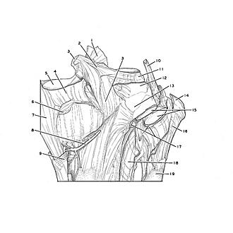

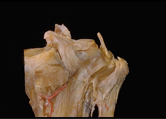
Dissection of knee
Cavity of right tibiofibular joint in relation to knee joint, posterior view
The posterior ligament of the head of the fibula has been divided. The fibula has been rotated forward to open the tibiofibular articulation.
- Anterior cruciate ligament (cut off)
- Posterior meniscofemoral ligament (ligament of Wrisberg)
- Posterior cruciate ligament (cut off)
- Medial meniscus
- Medial condyle of tibia (superior articular surface)
- Articular capsule (cut off)
- Tibial collateral ligament
- Medial inferior genicular artery
- Tendon of insertion of sartorius muscle
- Collateral ligament of fibula
- Lateral meniscus (pointer on surface for tendon of popliteus muscle)
- Lateral condyle of tibia (superior articular surface)
- Subpopliteal recess
- Popliteal arcuate ligament
- Upper pointer: Articular surface of fibula Lower pointer: Articular surface of head of fibula
- Head of fibula
- Posterior head of fibula ligament (divided)
- Interosseous membrane of leg
- Body of fibula