
Bassett Collection of Stereoscopic Images of Human Anatomy
Dissection of knee
Interior of right knee joint, posterior view showing capsular ligaments and internal structures
Image #191-2
KEYWORDS: Bones joints cartilage, Knee, Muscles and tendons.
Creative Commons
Stanford holds the copyright to the David L. Bassett anatomical images and has assigned Creative Commons license Attribution-Share Alike 4.0 International to all of the images.

For additional information regarding use and permissions, please contact the Medical History Center.
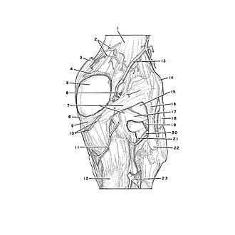
Dissection of knee
Interior of right knee joint, posterior view showing capsular ligaments and internal structures
Windows have been cut in the joint capsule posterior to the femoral and tibial condyles to expose the interior of the joint and at the same time to preserve the major capsular ligaments posteriorly.
- Popliteal femoral surface
- Medial superior genicular arteries
- Adductor magnus muscle (tendon of insertion)
- Articular capsule genus (cut across)
- Medial condyle of femur (covered by articular cartilage)
- Medial genicular artery (origin in common with lateral superior genicular artery)
- Upper pointer: Oblique popliteal ligament Lower pointer: Posterior meniscofemoral ligament
- Collateral ligament of tibia
- Medial meniscus (pointer indicates area of fusion with articular capsule)
- Semimembranosus muscle (tendon of insertion, cut off)
- Medial inferior genicular artery
- Body of tibia
- Lateral superior genicular artery
- Lateral epicondyle of femur
- Lateral condyle of femur (visible through opening in joint capsule)
- Popliteal arcuate ligament
- Lateral lemniscus
- Collateral ligament of fibula
- Popliteus muscle (tendon of origin, cut off)
- Lateral inferior genicular artery
- Subpopliteal recess
- Apex of fibula
- Anterior tibial artery