Bassett Collection of Stereoscopic Images of Human Anatomy
Dissection of knee
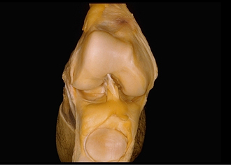
Interior of right knee joint, anterior view
Image #190-4
KEYWORDS: Bones joints cartilage, Knee, Muscles and tendons.
Creative Commons
Stanford holds the copyright to the David L. Bassett anatomical images and has assigned Creative Commons license Attribution-Share Alike 4.0 International to all of the images.

For additional information regarding use and permissions, please contact the Medical History Center.


Dissection of knee
Interior of right knee joint, anterior view
The joint capsule has been widely incised to permit the downward reflection of the quadriceps tendon, patella and patellar ligament.
- Suprapatellar bursa
- Lateral condyle of femur
- Lateral meniscus
- Infrapatellar fat body
- Patellar articular surface
- Quadriceps femoris muscle (reflected)
- Synovial membrane
- Articular capsule
- Intercondylar fossa
- Medial condyle of femur
- Infrapatellar synovial fold
- Medial meniscus
- Collateral ligament of tibia
- Alar folds