
Bassett Collection of Stereoscopic Images of Human Anatomy
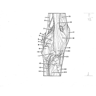
Dissection of knee
Capsule, ligaments and arteries of right knee, lateral view
Image #190-1
KEYWORDS: Knee, Muscles and tendons, Vasculature.
Creative Commons
Stanford holds the copyright to the David L. Bassett anatomical images and has assigned Creative Commons license Attribution-Share Alike 4.0 International to all of the images.

For additional information regarding use and permissions, please contact the Medical History Center.

Dissection of knee
Capsule, ligaments and arteries of right knee, lateral view
- Popliteal artery
- Muscular branch of popliteal artery
- Lateral superior genicular artery
- Medial condyle of femur (in background)
- Lateral condyle of femur (covered by ligaments)
- Semitendinosus muscle (tendon of insertion)
- Collateral ligament of fibula
- Popli teal arcuate ligament
- Biceps femoris muscle (tendon of insertion)
- Head of fibula
- Anterior head of fibula ligament
- Anterior tibial recurrent artery
- Anterior tibial artery
- Body of fibula
- Vastus lateralis muscle
- Iliotibial tract
- Patella
- Lateral patellar retinaculum
- Patellar ligament
- Lateral condyle of tibia
- Tibial tuberosity
- Body of tibia
- Interosseous membrane of leg