Bassett Collection of Stereoscopic Images of Human Anatomy
Exploration of those parts of the brain supplied by the posterior cerebral artery
Inferior and posterior horns of lateral ventricle
Image #19-6
KEYWORDS: Brain, Occipital lobe, Telencephalon, Temporal lobe, Ventricules.
Creative Commons
Stanford holds the copyright to the David L. Bassett anatomical images and has assigned Creative Commons license Attribution-Share Alike 4.0 International to all of the images.

For additional information regarding use and permissions, please contact the Medical History Center.
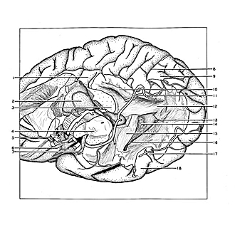
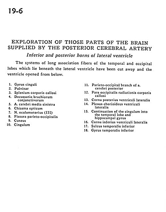
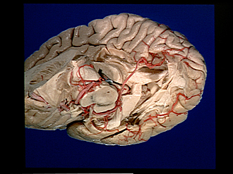
Exploration of those parts of the brain supplied by the posterior cerebral artery
Inferior and posterior horns of lateral ventricle
The systems of long association fibers of the temporal and occipital lobes which lie beneath the lateral ventricle have been cut away and the ventricle opened from below.
- Cingulate gyrus
- Pulvinar
- Corpus callosum (splenium)
- Decussation brachium conjunctivum (superior cerebeilar peduncle)
- Middle cerebral artery left
- Optic chiasm
- Oculomotor nerve (III)
- Parieto-occipital fissure
- Cuneus
- Cingulum
- Parieto-occipital branch of posterior cerebral artery
- Occipital part of corpus callosum radiations
- Posterior horn lateral ventricle
- Choroid plexus lateral ventricle
- Continuation of the cingulum into the temporal lobe and hippocampal gyrus
- Inferior horn of lateral ventricle
- Inferior temporal sulcus
- Inferior temporal gyrus