Bassett Collection of Stereoscopic Images of Human Anatomy
Exploration of those parts of the brain supplied by the posterior cerebral artery
Occipital radiation of corpus callosum; pulvinar
Image #19-5
KEYWORDS: Brain, Diencephalon, Occipital lobe, Telencephalon.
Creative Commons
Stanford holds the copyright to the David L. Bassett anatomical images and has assigned Creative Commons license Attribution-Share Alike 4.0 International to all of the images.

For additional information regarding use and permissions, please contact the Medical History Center.
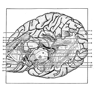
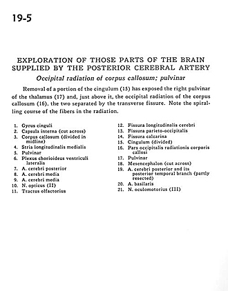
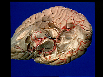
Exploration of those parts of the brain supplied by the posterior cerebral artery
Occipital radiation of corpus callosum; pulvinar
Removal of a portion of the cingulum (15) has exposed the right pulvinar of the thalamus (17) and, just above it, the occipital radiation of the corpus callosum (16), the two separated by the transverse fissure. Note the spiralling course of the fibers in the radiation.
- Cingulate gyrus
- Internal capsule (cut across )
- Corpus callosum (divided in midline)
- Medial longitudinal stria
- Pulvinar
- Choroid plexus lateral ventricle
- Posterior cerebral artery
- Middle cerebral artery
- Middle cerebral artery
- Optic nerve (II)
- Olfactory tract
- Longitudinal fissure (cerebral)
- Parieto-occipital fissure
- Calcarine fissure
- Cingulum (divided)
- Occipital part radiations of corpus callosum
- Pulvinar
- Mesencephalon (cut across)
- Posterior cerebral artery and its posterior temporal branch (partly resected)
- Basilar artery
- Oculomotor nerve (III)