Bassett Collection of Stereoscopic Images of Human Anatomy
Dissection of knee
Superficial vessels and nerves of left knee; patellar retinaculum; medial view
Image #189-6
KEYWORDS: Bones joints cartilage, Fascia, Knee, Muscles and tendons, Peripheral nervous system, Vasculature.
Creative Commons
Stanford holds the copyright to the David L. Bassett anatomical images and has assigned Creative Commons license Attribution-Share Alike 4.0 International to all of the images.

For additional information regarding use and permissions, please contact the Medical History Center.
Dissection of knee
Superficial vessels and nerves of left knee; patellar retinaculum; medial view
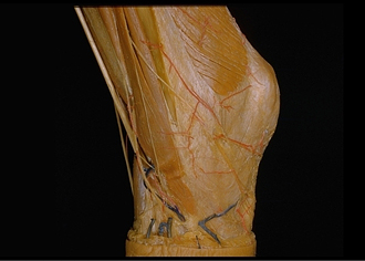
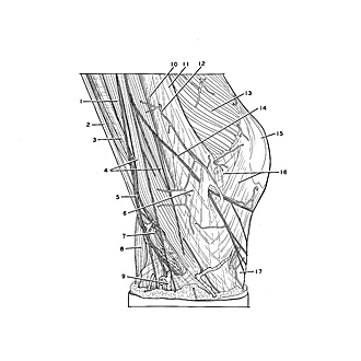
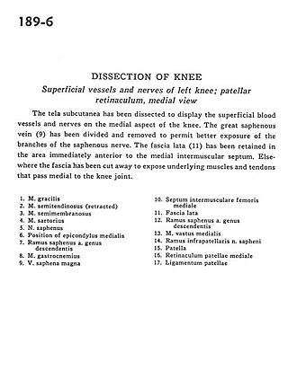
The tela subcutanea has been dissected to display the superficial blood vessels and nerves on the medial aspect of the knee. The great saphenous vein (9) has been divided and removed to permit better exposure of the branches of the saphenous nerve. The fascia lata (11) has been retained in the area immediately anterior to the medial intermuscular septum. Elsewhere the fascia has been cut away to expose underlying muscles and tendons that pass medial to the knee joint.
- Gracilis muscle
- Semitendinosus muscle (retracted)
- Semimembranosus muscle
- Sartorius muscle
- Saphenous nerve
- Position of medial epicondyle
- Saphenous branch of descending genicular artery
- Gastrocnemius muscle
- Greater saphenous vein
- Medial femoral intermuscular septum
- Fascia lata
- Saphenous branch of descending genicular artery
- Vastus medialis muscle
- Infrapatellar branch of saphenous nerve
- Patella
- Medial patellar retinaculum
- Patellar ligament