Bassett Collection of Stereoscopic Images of Human Anatomy
Dissection of knee
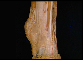
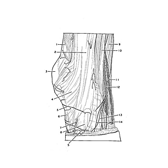
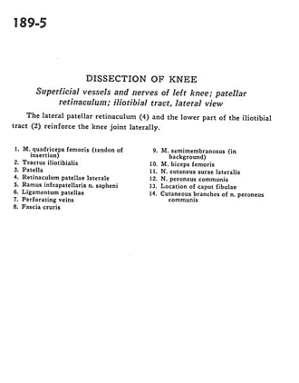
Superficial vessels and nerves of left knee; patellar retinaculum; iliotibial tract, lateral view
Image #189-5
KEYWORDS: Bones joints cartilage, Knee, Peripheral nervous system, Vasculature.
Creative Commons
Stanford holds the copyright to the David L. Bassett anatomical images and has assigned Creative Commons license Attribution-Share Alike 4.0 International to all of the images.

For additional information regarding use and permissions, please contact the Medical History Center.
Dissection of knee
Superficial vessels and nerves of left knee; patellar retinaculum; iliotibial tract, lateral view
The lateral patellar retinaculum (4) and the lower part of the iliotibial tract (2) reinforce the knee joint laterally.
- Quadriceps femoris muscle (tendon of insertion)
- Iliotibial tract
- Patella
- Lateral patellar retinaculum
- Infrapatellar branch of saphenous nerve
- Patellar ligament
- Perforating veins
- Crural fascia
- Semimembranosus muscle (in background)
- Biceps femoris muscle
- Lateral cutaneous sural nerve
- Common peroneal nerve
- Location of head of fibula
- Cutaneous branches of common peroneal nerve