
Bassett Collection of Stereoscopic Images of Human Anatomy
Dissection of anterior and medial aspects of thigh
Quadriceps femoris muscle (continued).
Image #188-7
KEYWORDS: Muscles and tendons, Thigh.
Creative Commons
Stanford holds the copyright to the David L. Bassett anatomical images and has assigned Creative Commons license Attribution-Share Alike 4.0 International to all of the images.

For additional information regarding use and permissions, please contact the Medical History Center.

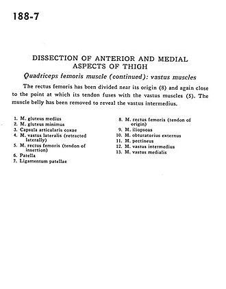
Dissection of anterior and medial aspects of thigh
Quadriceps femoris muscle (continued).
The rectus femoris has been divided near its origin (8) and again close to the point at which its tendon fuses with the vastus muscles (5). The muscle belly has been removed to reveal the vastus intermedius.
- Gluteus medius muscle
- Gluteus minimus muscle
- Hip articular capsule
- Vastus lateralis muscle (retracted laterally)
- Rectus lemons muscle (tendon of insertion)
- Patella
- Patellar ligament
- Rectus femoris muscle (tendon of origin)
- Iliopsoas muscle
- Obturator externus muscle
- Pectineus muscle
- Vastus intermedius muscle
- Vastus medialis muscle