Bassett Collection of Stereoscopic Images of Human Anatomy
Dissection of anterior and medial aspects of thigh
Obturator externus muscle
Image #188-4
KEYWORDS: Muscles and tendons, Thigh.
Creative Commons
Stanford holds the copyright to the David L. Bassett anatomical images and has assigned Creative Commons license Attribution-Share Alike 4.0 International to all of the images.

For additional information regarding use and permissions, please contact the Medical History Center.
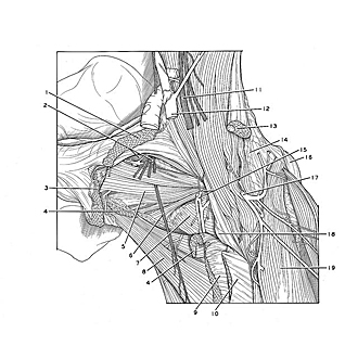
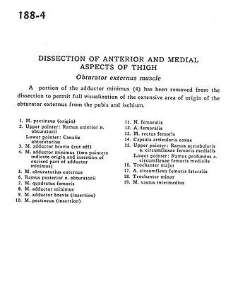
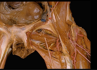
Dissection of anterior and medial aspects of thigh
Obturator externus muscle
A portion of the adductor minimus (4) has been removed from the dissection to permit full visualization of the extensive area of origin of the obturator externus from the pubis and ischium.
- Pectineus muscle (origin)
- Upper pointer: Anterior branch of obturator nerve Lower pointer: Obturator canal
- Adductor brevis muscle (cut off)
- Adductor minimus muscle (two pointers indicate origin and insertion of excised part of adductor minimus)
- Obturator externus muscle
- Posterior branch of obturator nerve
- Quadratus femoris muscle
- Adductor minimus muscle
- Adductor brevis muscle (insertion)
- Pectineus muscle (insertion)
- Femoral nerve
- Femoral artery
- Rectus femoris muscle
- Hip articular capsule
- Upper pointer: Acetabular branch of medial circumflex femoral artery Lower pointer: Deep branch of medial circumflex femoral artery
- Greater trochanter
- Lateral femoral circumflex artery
- Lesser trochanter
- Vastus intermedius muscle