
Bassett Collection of Stereoscopic Images of Human Anatomy
Dissection of anterior and medial aspects of thigh
Posterior branch of obturator nerve; innervation of adductor magnus by obturator nerve
Image #188-3
KEYWORDS: Muscles and tendons, Peripheral nervous system, Thigh.
Creative Commons
Stanford holds the copyright to the David L. Bassett anatomical images and has assigned Creative Commons license Attribution-Share Alike 4.0 International to all of the images.

For additional information regarding use and permissions, please contact the Medical History Center.
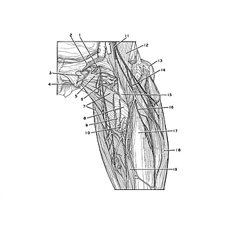
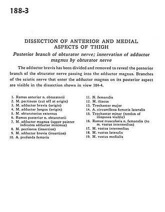
Dissection of anterior and medial aspects of thigh
Posterior branch of obturator nerve; innervation of adductor magnus by obturator nerve
The adductor brevis has been divided and removed to reveal the posterior branch of the obturator nerve passing into the adductor magnus. Branches of the sciatic nerve that enter the adductor magnus on its posterior aspect are visible in the dissection shown in view 184-4.
- Anterior branch of obturator nerve
- Pectineus muscle (cut off at origin)
- Adductor brevis muscle (origin)
- Adductor longus muscle (origin)
- Obturator externus muscle
- Posterior branch of obturator nerve
- Adductor magnus muscle (upper pointer indicates adductor minimus)
- Pectineus muscle (insertion)
- Adductor brevis muscle (insertion)
- Deep femoral artery
- Femoral nerve
- Iliacus muscle
- Greater trochanter
- Lateral femoral circumflex artery
- Lesser trochanter (tendon of iliopsoas visible)
- Muscular branch of femoral nerve (to vastus intermedius muscle)
- Vastus intermedius muscle
- Vastus lateralis muscle
- Vastus medialis muscle