Bassett Collection of Stereoscopic Images of Human Anatomy
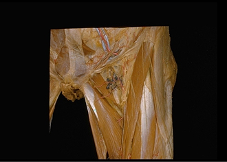
Dissection of anterior and medial aspects of thigh
Left femoral triangle with superficial nerves and vessels retained, anterior view
Image #186-1
KEYWORDS: Fascia, Muscles and tendons, Peripheral nervous system, Thigh, Vasculature.
Creative Commons
Stanford holds the copyright to the David L. Bassett anatomical images and has assigned Creative Commons license Attribution-Share Alike 4.0 International to all of the images.

For additional information regarding use and permissions, please contact the Medical History Center.


Dissection of anterior and medial aspects of thigh
Left femoral triangle with superficial nerves and vessels retained, anterior view
The fascia lata has been retained in the area surrounding the saphenous opening. Superficial inguinal lymph nodes have beem cut away althrough the lymphatic vessels (7) that drain these nodes and pass through the cribriform fascia (8) have been preserved.
- Transversalis fascia
- Inguinal ligament (slightly elevated)
- Iliopubic tract
- Inferior epigastric veins
- Inferior epigastric artery
- Round ligament of uterus
- Lymphatic vessel (note other lymphatic vessels in the area)
- Upper pointer: Cribriform fascia Lower pointer: Greater saphenous vein (approaching ovalis fossa of fascia lata)
- External pudendal arteries
- Pectineus muscle
- Fascia lata
- Adductor longus muscle
- Cutaneous branches of anterior femoral nerve
- Gracilis muscle
- Lateral femoral cutaneous nerve
- Tensor fasciae latae muscle
- Femoral branch of genitofemoral nerve
- Sartorius muscle
- Margo falciformis hiatus sapheni
- Rectus femoris muscle