Bassett Collection of Stereoscopic Images of Human Anatomy
Dissection of anterior and medial aspects of thigh
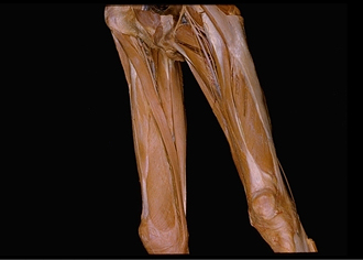
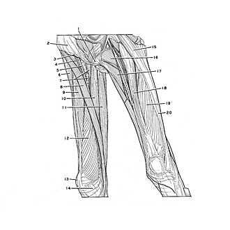
Muscles of right thigh, anteromedial view
Image #185-6
KEYWORDS: Muscles and tendons, Thigh.
Creative Commons
Stanford holds the copyright to the David L. Bassett anatomical images and has assigned Creative Commons license Attribution-Share Alike 4.0 International to all of the images.

For additional information regarding use and permissions, please contact the Medical History Center.

Dissection of anterior and medial aspects of thigh
Muscles of right thigh, anteromedial view
The specimen has been turned so that the view is directed toward the anteromedial aspect of the right thigh. The muscles have been spread apart.
- Location of pubic symphysis
- Aponeurosis External oblique muscle
- Inguinal ligament
- Femoral nerve
- Femoral artery
- Tensor fasciae latae muscle
- Upper pointer: Pectineus muscle Lower pointer: Femoral vein
- Sartorius muscle
- Rectus femoris muscle
- Adductor longus muscle
- Gracilis muscle
- Vastus medialis muscle
- Patella
- Medial patellar retinaculum
- Tensor fasciae latae muscle
- Pectineus muscle
- Adductor longus muscle
- Rectus femoris muscle
- Vastus lateralis muscle
- Iliotibial tract