
Bassett Collection of Stereoscopic Images of Human Anatomy
Dissection of anterior and medial aspects of thigh
Muscles of left thigh, anterior view
Image #185-5
KEYWORDS: Fascia, Muscles and tendons, Thigh.
Creative Commons
Stanford holds the copyright to the David L. Bassett anatomical images and has assigned Creative Commons license Attribution-Share Alike 4.0 International to all of the images.

For additional information regarding use and permissions, please contact the Medical History Center.

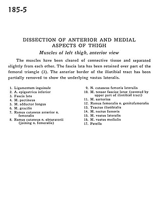
Dissection of anterior and medial aspects of thigh
Muscles of left thigh, anterior view
The muscles have been cleared of connective tissue and separated slightly from each other. The fascia lata has been retained over part of the femoral triangle (3). The anterior border of the iliotibial tract has been partially removed to show the underlying vastus lateralis.
- Inguinal ligament
- Inferior epigastric artery
- Fascia lata
- Pectineus muscle
- Adductor longus muscle
- Gracilis muscle
- Anterior cutaneous branch of femoral nerve
- Cutaneous branch of obturator nerve (joining femoral nerve)
- Lateral femoral cutaneous nerve
- Tensor fasciae latae muscle (covered by upper part of iliotibial tract)
- Sartorius muscle
- Femoral branch of genitofemoral nerve
- Iliotibial tract
- Rectus femoris muscle
- Vastus lateralis muscle
- Vastus medialis muscle
- Patella