Bassett Collection of Stereoscopic Images of Human Anatomy
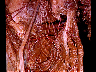
Dissection of posterior aspect of left thigh
Adductor minimus muscle
Image #184-5
KEYWORDS: Muscles and tendons, Thigh.
Creative Commons
Stanford holds the copyright to the David L. Bassett anatomical images and has assigned Creative Commons license Attribution-Share Alike 4.0 International to all of the images.

For additional information regarding use and permissions, please contact the Medical History Center.
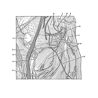

Dissection of posterior aspect of left thigh
Adductor minimus muscle
The sciatic nerve has been retracted laterally and the adductor magnus has been slightly deflected medially to reveal the adductor minimus. This muscle is often fused with the adductor magnus and is usually described as a part of it.
- Trochanter tertius
- Gluteus maximus muscle (reflected)
- Sciatic nerve
- Perforating artery
- Perforating artery
- Quadratus femoris muscle
- Origin of biceps femoris muscle
- Origin of semimembranosus muscle
- Origin of semitendinosus muscle
- Medial femoral circumflex artery
- Adductor magnus muscle
- Adductor minimus muscle
- Muscular branch of sciatic nerve (to adductor magnus muscle)