Bassett Collection of Stereoscopic Images of Human Anatomy
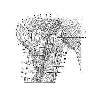
Dissection of posterior aspect of left thigh
Relations of sciatic nerve in upper part of thigh (continued)
Image #183-4
KEYWORDS: Peripheral nervous system, Thigh.
Creative Commons
Stanford holds the copyright to the David L. Bassett anatomical images and has assigned Creative Commons license Attribution-Share Alike 4.0 International to all of the images.

For additional information regarding use and permissions, please contact the Medical History Center.

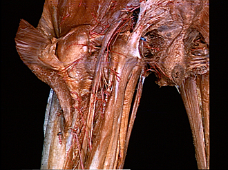
Dissection of posterior aspect of left thigh
Relations of sciatic nerve in upper part of thigh (continued)
The sciatic nerve has been exposed from the greater sciatic foramen downward into the thigh. The relations of the nerve to the ischial tuberosity (19) and greater trochanter (6) are shown. Several arteries (1, 2, 7, 14, 21) ramify within the area of the dissection. These vessels, together with branches of the lateral circumflex femoral artery (not shown) form the cruciate anastomosis.
- Inferior gluteal artery
- Artery supplying sciatic nerve
- Piriform muscle
- Obturator internus muscle
- Gluteus medius muscle
- Greater trochanter
- Medial femoral circumflex artery (ascending branch)
- Fascia lata (between gluteus maximus muscle and tensor fasciae latae muscle)
- Trochanteric bursa of gluteus maximus
- Gluteus maximus muscle (reflected)
- Quadratus femoris muscle
- Adductor minimus muscle
- Sciatic nerve
- Perforating arteries
- Lateral intermuscular septum
- Adductor magnus muscle
- Short head of biceps femoris muscle
- Sacrotuberous ligament
- Ischial tuberosity
- Muscular branch of sciatic nerve (to hamstring muscles)
- Medial femoral circumflex artery (deep branch)
- Adductor magnus muscle
- Gracilis muscle
- Semitendinosus muscle
- Long head of biceps femoris