Bassett Collection of Stereoscopic Images of Human Anatomy
Dissection of posterior aspect of left thigh
Popliteal fossa, close-up view
Image #183-1
KEYWORDS: Muscles and tendons, Peripheral nervous system, Thigh.
Creative Commons
Stanford holds the copyright to the David L. Bassett anatomical images and has assigned Creative Commons license Attribution-Share Alike 4.0 International to all of the images.

For additional information regarding use and permissions, please contact the Medical History Center.
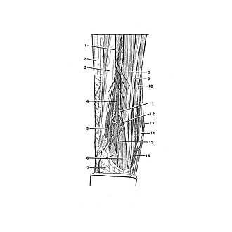
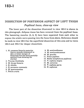
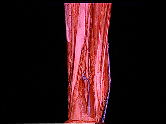
Dissection of posterior aspect of left thigh
Popliteal fossa, close-up view
The lower part of the dissection illustrated in view 182-6 is shown in this photograph. Adipose tissue has been removed from the popliteal fossa. The hamstring muscles (3, 8, 9) have been separated from each other to expose the sciatic nerve passing into the fossa from above. Reference should be made to view 182-5 for the superficial dissection of this area and to views 183-5 and 190-2 for deeper dissections.
- Posterior femoral cutaneous nerve (nerve extends downward to skin at lower margin of dissection but has been partially omitted from drawing for clarity)
- Iliotibial tract
- Biceps femoris muscle
- Tibial nerve
- Common peroneal nerve
- Gastrocnemius muscle
- Crural fascia
- Semitendinosus muscle
- Semimembranosus muscle
- Gracilis muscle
- Popliteal fossa
- Popliteal lymph node
- Greater saphenous vein
- Sartorius muscle
- Lesser saphenous vein
- Saphenous nerve