
Bassett Collection of Stereoscopic Images of Human Anatomy
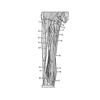
Dissection of posterior aspect of left thigh
Posterior muscles of thigh
Image #182-6
KEYWORDS: Muscles and tendons, Thigh.
Creative Commons
Stanford holds the copyright to the David L. Bassett anatomical images and has assigned Creative Commons license Attribution-Share Alike 4.0 International to all of the images.

For additional information regarding use and permissions, please contact the Medical History Center.
Dissection of posterior aspect of left thigh
Posterior muscles of thigh
The fascia lata has been removed and the underlying muscles have been cleaned of connective tissue and slightly separated from each other.
- Gluteus maximus muscle
- Iliotibial tract
- Posterior femoral cutaneous nerve
- Long head of biceps femoris muscle
- Short head of biceps femoris muscle
- Tibial nerve
- Common peroneal nerve
- Lesser saphenous.vein
- Ischial tuberosity
- Perineal branch of posterior femoral cutaneous nerve
- Adductor magnus muscle
- Semitendinosus muscle
- Semimembranosus muscle
- Gracilis muscle
- Sartorius muscle
- Greater saphenous vein
- muscle gastrocnemius