
Bassett Collection of Stereoscopic Images of Human Anatomy
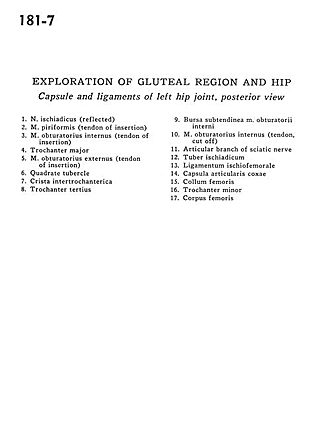
Exploration of gluteal region and hip
Capsule and ligament of left hip joint, posterior view
Image #181-7
KEYWORDS: Bones joints cartilage, Muscles and tendons.
Creative Commons
Stanford holds the copyright to the David L. Bassett anatomical images and has assigned Creative Commons license Attribution-Share Alike 4.0 International to all of the images.

For additional information regarding use and permissions, please contact the Medical History Center.

Exploration of gluteal region and hip
Capsule and ligament of left hip joint, posterior view
- Sciatic nerve (reflected)
- Piriform muscle (tendon of insertion)
- Obturator internus muscle (tendon of insertion)
- Greater trochanter
- Obturator externus muscle (tendon of insertion)
- Quadrate tubercle
- Intertrochanteric crest
- Trochanter tertius
- Subtendinous bursa of obturator internus muscle
- Obturator internus muscle (tendon, cut off)
- Articular branch of sciatic nerve
- Ischial tuberosity
- Ischiofemoral ligament
- Flip articular capsule
- Neck of femur
- Lesser trochanter
- Body of femur