Bassett Collection of Stereoscopic Images of Human Anatomy
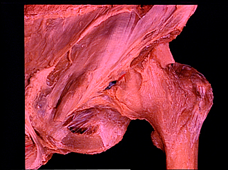
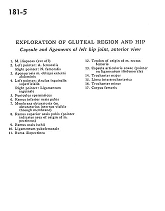
Exploration of gluteal region and hip
Capsule and ligaments of left hip joint, anterior view
Image #181-5
KEYWORDS: Bones joints cartilage, Muscles and tendons.
Creative Commons
Stanford holds the copyright to the David L. Bassett anatomical images and has assigned Creative Commons license Attribution-Share Alike 4.0 International to all of the images.

For additional information regarding use and permissions, please contact the Medical History Center.
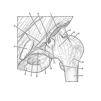
Exploration of gluteal region and hip
Capsule and ligaments of left hip joint, anterior view
- Iliopsoas muscle (cut off)
- Left pointer: Femoral artery Right pointer: Femoral nerve
- Aponeurosis of external oblique muscle
- Left pointer: Superficial inguinal ring Right pointer: Inguinal ligament
- Spermatic cord
- Inferior pubic ramus
- Obturator membrane (Obturator internus muscle visible through membrane)
- Superior pubic ramus (pointer indicates area of origin of pectineus muscle)
- Ischial ramus
- Pubofemoral ligament
- Iliopectineal bursa
- Tendon of origin of rectus femoris muscle
- Hip articular capsule (pointer on iliofemoral ligament)
- Greater trochanter
- Intertrochanteric line
- Lesser trochanter
- Body of femur