Bassett Collection of Stereoscopic Images of Human Anatomy
Exploration of gluteal region and hip
Relation of gluteus minimus to capsule of hip joint; nerve to hip joint
Image #180-7
KEYWORDS: Bones joints cartilage, Muscles and tendons, Peripheral nervous system.
Creative Commons
Stanford holds the copyright to the David L. Bassett anatomical images and has assigned Creative Commons license Attribution-Share Alike 4.0 International to all of the images.

For additional information regarding use and permissions, please contact the Medical History Center.

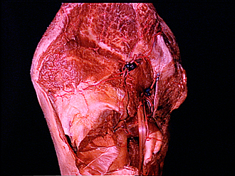
Exploration of gluteal region and hip
Relation of gluteus minimus to capsule of hip joint; nerve to hip joint
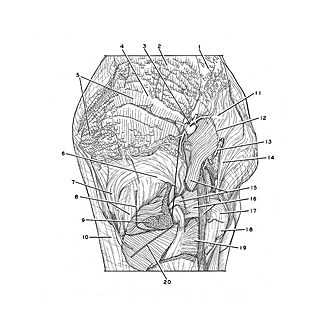
The gluteus minimus has been reflected downward to reveal the capsule of the hip joint. A branch of the superior gluteal nerve to the capsule is stretched around the retracted muscle.
- Area of origin of Gluteus maximus muscle
- Area of origin of Gluteus medius muscle
- Superior gluteal artery
- Location of Anterior gluteal line
- Area of origin of Gluteus minimus muscle
- Acetabular margin (visible through capsule of hip joint)
- Reflected head of origin of Rectus femoris muscle
- Upper pointer: Branch of Superior gluteal nerve to hip joint capsule Lower pointer: Hip articular capsule
- Gluteus minimus muscle (reflected)
- Iliotibial tract (covering tensor fasciae latae muscle)
- Posterior inferior iliac spine
- Margin of Greater sciatic foramen
- Inferior gluteal artery
- Sacrotuberous ligament
- Piriform muscle (divided)
- Upper pointer: Obturator internus muscle Lower pointer: Sciatic nerve
- Ischial tuberosity
- Biceps femoris muscle (origin of long head)
- Quadratus femoris muscle
- Nerve to tensor fasciae latae muscle (cut off)