Bassett Collection of Stereoscopic Images of Human Anatomy
Exploration of the brain from the medial aspect
Claustrum, inferior occipitofrontal fasciculus, amygdaloid nucleus and inferior horn of lateral ventricle
Image #18-6
KEYWORDS: Brain, Telencephalon, Ventricules.
Creative Commons
Stanford holds the copyright to the David L. Bassett anatomical images and has assigned Creative Commons license Attribution-Share Alike 4.0 International to all of the images.

For additional information regarding use and permissions, please contact the Medical History Center.


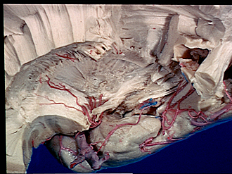
Exploration of the brain from the medial aspect
Claustrum, inferior occipitofrontal fasciculus, amygdaloid nucleus and inferior horn of lateral ventricle
The thin gray lamina of the claustrum has been exposed by removal of part of the external capsule. More posteriorly in this area the medial fibers of the inferior occipitofrontal fasciculus (11) are also uncovered. The choroidal fissure is opened and the substantia innominata and part of the amygdaloid nucleus removed so that the inferior horn of the lateral ventricle can be seen.
- External capsule
- Claustrum
- Anterior commissure
- "Substantia innominata" and amygdaloid nucleus (dissected)
- Middle cerebral artery
- Temporal pole
- Superior longitudinal fasciculus
- Corona radiata
- Choroid plexus lateral ventricle
- Fornix (crus) (cut across)
- Inferior occipitofrontal fasciculus
- Sublenticular part of internal capsule (cut across)
- Stria terminalis
- Fimbria of hippocampus
- Inferior horn of lateral ventricle
- Choroidal artery (anterior)