Bassett Collection of Stereoscopic Images of Human Anatomy
Exploration of gluteal region and hip
Deep relations of gluteus maximus, left posterolateral view
Image #179-7
KEYWORDS: Muscles and tendons, Peripheral nervous system, Vasculature.
Creative Commons
Stanford holds the copyright to the David L. Bassett anatomical images and has assigned Creative Commons license Attribution-Share Alike 4.0 International to all of the images.

For additional information regarding use and permissions, please contact the Medical History Center.
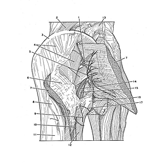
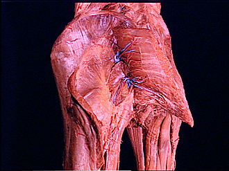
Exploration of gluteal region and hip
Deep relations of gluteus maximus, left posterolateral view
The gluteus maximus has been divided transversely across its midportion and the ends of the muscle have been reflected medially and laterally. The medial part of the lamina of fascia lata underlying the muscle has been removed. The gluteus medius and piriformis muscles have been exposed together with the sciatic nerve and branches of the gluteal vessels and nerves. The medial area of this dissection is shown in detail in the following view.
- Superior gluteal artery (accompanied by superior gluteal vein)
- Quadratus lumborum muscle
- Iliac crest
- Gluteus medius (partially covered by fascia lata)
- Upper pointer: Inferior gluteal nerve Lower pointer: piriform muscle
- Fascia lata (deep to gluteus maximus)
- Gluteus maximus muscle (divided and reflected)
- Location of greater trochanter
- Inferior cluneal nerve (cut off)
- Posterior femoral cutaneous nerve
- Iliotibial tract
- Biceps femoris muscle
- Vertebral spinous process of L. IV
- Sciatic nerve
- Artery supplying sciatic nerve
- Ischial tuberosity
- Perineal branch of posterior femoral cutaneous nerve