Bassett Collection of Stereoscopic Images of Human Anatomy
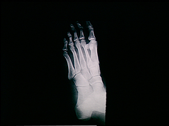
Radiography
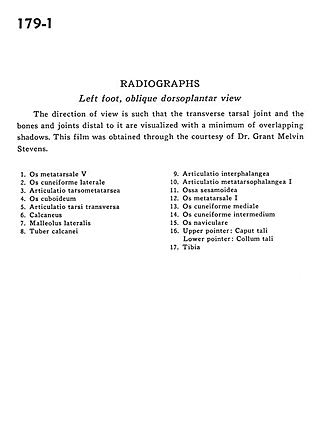
Left foot, oblique dorsoplantar view
Image #179-1
KEYWORDS: Bones joints cartilage, Foot and toes.
Creative Commons
Stanford holds the copyright to the David L. Bassett anatomical images and has assigned Creative Commons license Attribution-Share Alike 4.0 International to all of the images.

For additional information regarding use and permissions, please contact the Medical History Center.
Radiography
Left foot, oblique dorsoplantar view
The direction of view is such that the transverse tarsal joint and the bones and joints distal to it are visualized with a minimum of overlapping shadows. This film was obtained through the courtesy of Dr. Grant Melvin Stevens.
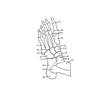
- 5th metatarsal bone
- Lateral cuneiform bone
- Tarsometatarsal articulation
- Cuboid bone
- Transverse tarsal articulation
- Calcaneus
- Lateral malleolus
- Tuberosity of calcaneus
- Interphalangeal articulation
- 1st metatarsophalangeal articulations
- Sesamoid bone
- Metatarsal bone
- Medial cuneiform bone
- Intermediate cuneiform bone
- Navicular bone
- Upper pointer: Head of talus Lower pointer: Neck of talus
- Tibia