Bassett Collection of Stereoscopic Images of Human Anatomy
Osteology
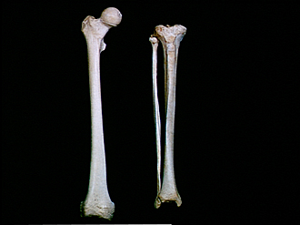
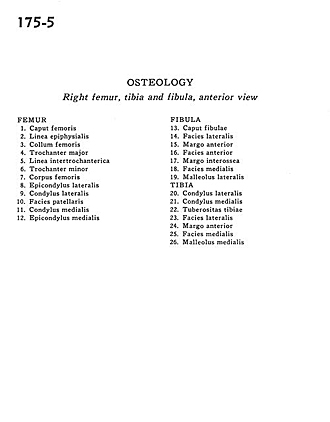
Right femur, tibia and fibula, anterior view
Image #175-5
KEYWORDS: Bones joints cartilage, Leg, Thigh.
Creative Commons
Stanford holds the copyright to the David L. Bassett anatomical images and has assigned Creative Commons license Attribution-Share Alike 4.0 International to all of the images.

For additional information regarding use and permissions, please contact the Medical History Center.
Osteology
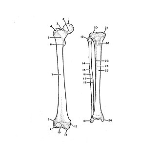
Right femur, tibia and fibula, anterior view
- Femur
- Head of femur
- Epiphysial line
- Neck of femur
- Greater trochanter
- Intertrochanteric line
- Lesser trochanter
- Body of femur
- Lateral epicondyle
- Lateral condyle
- Patellar surface
- Medial condyle
- Medial epicondyle
- Fibula
- Head of fibula
- Lateral surface
- Anterior border
- Anterior surface
- Interosseous border
- Medial surface
- Lateral malleolus
- Tibia
- Lateral condyle
- Medial condyle
- Tibial tuberosity
- Lateral surface
- Anterior border
- Medial surface
- Medial malleolus