
Bassett Collection of Stereoscopic Images of Human Anatomy
Dissection of muscles of male perineum and pelvic diaphragm
Pelvic diaphragm viewed from above
Image #174-7
KEYWORDS: Muscles and tendons.
Creative Commons
Stanford holds the copyright to the David L. Bassett anatomical images and has assigned Creative Commons license Attribution-Share Alike 4.0 International to all of the images.

For additional information regarding use and permissions, please contact the Medical History Center.
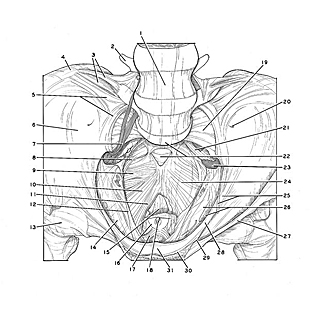
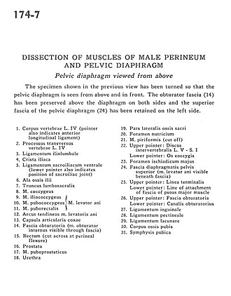
Dissection of muscles of male perineum and pelvic diaphragm
Pelvic diaphragm viewed from above
The specimen shown in the previous view has been turned so that the pelvic diaphragm is seen from above and in front. The obturator fascia (14) has been preserved above the diaphragm on both sides and the superior fascia of the pelvic diaphragm (24) has been retained on the left side.
- Body of vertebra L. IV (pointer also indicates anterior longitudinal ligament)
- Transverse process vertebra L. IV
- Iliolumbar ligament
- Iliac crest
- Ventral sacroiliac ligament (lower pointer also indicates position of sacroiliac joint)
- Ala of ilium
- Lumbosacral trunk
- Coccygeus muscle
- Iliococcygeus muscle
- Pubococcygeus muscle
- Puborectalis muscle (9-11 make up the levator ani muscle)
- Tendinous arch of levator ani muscle
- Joint capsule of coccyx
- Obturator fascia (obturator internus muscle visible through fascia)
- Rectum (cut across at perineal flexure)
- Prostate
- Puboprostatic muscle
- Urethra
- Lateral part of sacrum
- Nutritive foramen
- Piriform muscle (cut off)
- Upper pointer: Intervertebral disc L. V - S. I Lower pointer: Coccyx
- Greater sciatic foramen
- Superior fascia of pelvic diaphragm (Levator ani muscle visible beneath fascia)
- Upper pointer: Linea terminalis Lower pointer: Line of attachment of fascia of psoas major muscle
- Upper pointer: Obturator fascia Lower pointer: Obturator canal
- Inguinal ligament
- Pectineal ligament
- Lacunar ligament
- Body of pubic bone
- Pubic symphysis