
Bassett Collection of Stereoscopic Images of Human Anatomy
Dissection of muscles of male perineum and pelvic diaphragm
Inferior fascia of urogenital diaphragm (perineal membrane)
Image #174-3
KEYWORDS: Muscles and tendons.
Creative Commons
Stanford holds the copyright to the David L. Bassett anatomical images and has assigned Creative Commons license Attribution-Share Alike 4.0 International to all of the images.

For additional information regarding use and permissions, please contact the Medical History Center.
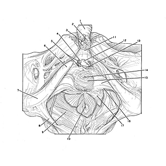
Dissection of muscles of male perineum and pelvic diaphragm
Inferior fascia of urogenital diaphragm (perineal membrane)
The penis has been transected opposite the pubic symphysis. The inferior fascia of the urogenital diaphragm has been preserved and the dorsal nerves, arteries, and vein of the penis are visible in relation to the anterior border of the diaphragm.
- Body of penis (covered by penile fascia)
- Corpus spongiosum of penis (pointer indicates cut end of urethra)
- Corpora cavernosa penis (joined by septum penis)
- Pubic arcuate ligament
- Upper pointer: Dorsal nerve of the penis Lower pointer: Dorsal artery of penis
- Inferior pubic ramus
- Ischial tuberosity
- Sacrotuberous ligament
- Levator ani muscle
- Anus
- Suspensory ligament of the penis
- Dorsal vein of the penis
- Transverse perineal ligament
- Urethra (cut off at junction of membranous and spongy parts)
- Left pointer: Sphincter muscle of urethra (visible through thin part of perineal membrane) Right pointer: Inferior fascia of urogenital diaphragm
- Posterior border of urogenital diaphragm
- Fibers of levator ani that insert in tissue alongside anus