
Bassett Collection of Stereoscopic Images of Human Anatomy
Dissection of male pelvis from a lateral approach
Piriformis muscle and nerve supply, medial view
Image #173-1
KEYWORDS: Muscles and tendons, Peripheral nervous system.
Creative Commons
Stanford holds the copyright to the David L. Bassett anatomical images and has assigned Creative Commons license Attribution-Share Alike 4.0 International to all of the images.

For additional information regarding use and permissions, please contact the Medical History Center.
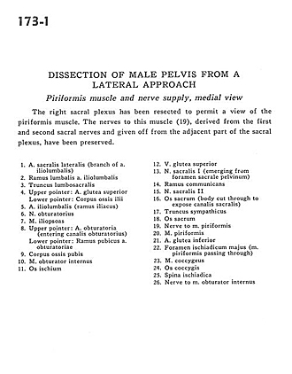
Dissection of male pelvis from a lateral approach
Piriformis muscle and nerve supply, medial view
The right sacral plexus has been resected to permit a view of the piriformis muscle. The nerves to this muscle (19), derived from the first and second sacral nerves and given off from the adjacent part of the sacral plexus, have been preserved.
- Lateral sacral artery (branch of iliolumbar artery)
- Lumbar branch iliolumbar artery
- Lumbosacral trunk
- Upper pointer: Superior gluteal artery Lower pointer: Body of ilium
- Iliolumbar artery (iliac branch of)
- Obturator nerve
- Iliopsoas muscle
- Upper pointer: Obturator artery (entering obturator canal) Lower pointer: Pubic branch of obturator artery
- Body of pubic bone
- Obturator internus muscle
- Ischium
- Superior gluteal vein
- Sacral nerve I (emerging from anterior (pelvic) sacral foramen)
- Ramus communicans
- Sacral nerve II
- Sacrum (body cut through to expose sacral canal)
- Sympathetic trunk
- Sacrum
- Nerve to piriform muscle
- Piriform muscle
- Inferior gluteal artery
- Greater sciatic foramen (piriform muscle passing through)
- Coccygeus muscle
- Coccyx
- Ischial spine
- Nerve to obturator internus muscle