Bassett Collection of Stereoscopic Images of Human Anatomy
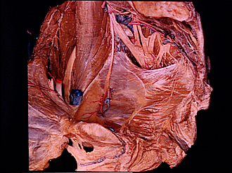
Dissection of male pelvis from a lateral approach
Sacral plexus and pelvic diaphragm, medial view with blood vessels removed
Image #172-6
KEYWORDS: Muscles and tendons, Vasculature.
Creative Commons
Stanford holds the copyright to the David L. Bassett anatomical images and has assigned Creative Commons license Attribution-Share Alike 4.0 International to all of the images.

For additional information regarding use and permissions, please contact the Medical History Center.

Dissection of male pelvis from a lateral approach
Sacral plexus and pelvic diaphragm, medial view with blood vessels removed
The arteries and veins have been removed from the central area of dissection to expose the sacral plexus as it passes through the greater sciatic foramen.
- Iliolumbar artery
- Psoas major muscle
- Iliacus muscle
- Obturator nerve
- Femoral nerve
- Linea terminalis
- Ductus deferens (cut off)
- External iliac artery and vein (passing downward deep to inguinal ligament)
- Obturator artery
- Pubic symphysis (sectioned)
- Obturator internus muscle (covered by obturator fascia)
- Tendinous arch pelvic fascia
- Urethra (opened)
- Urogenital diaphragm (dissected)
- Lumbosacral trunk
- Lateral sacral artery
- Superior gluteal artery
- Sympathetic trunk (pointer on ganglion)
- Left pointer: Greater sciatic foramen Right pointer: Sciatic nerve
- Sacrum (partially resected)
- Sacral nerve III (ventral ramus)
- Piriformis muscle
- Sacral nerve IV
- Upper pointer: Inferior gluteal artery Lower pointer: Internal pudendal artery
- Nerve to Levator ani muscle
- Coccygeus muscle
- Levator ani muscle
- Anal canal