Bassett Collection of Stereoscopic Images of Human Anatomy
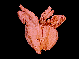
Dissection of male pelvis from a lateral approach
Interior of seminal vesicle, ejaculatory duct and ampulla of ductus deferens
Image #171-4
KEYWORDS:
Creative Commons
Stanford holds the copyright to the David L. Bassett anatomical images and has assigned Creative Commons license Attribution-Share Alike 4.0 International to all of the images.

For additional information regarding use and permissions, please contact the Medical History Center.
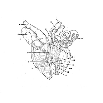
Dissection of male pelvis from a lateral approach
Interior of seminal vesicle, ejaculatory duct and ampulla of ductus deferens
The prostate shown in the preceding view has been removed from the body for separate dissection. The median lobe (7) has been removed to the left of the midline to gain access to the left ejaculatory duct. The ejaculatory duct has been opened and incisions have been continued to open the left seminal vesicle and the ampulla of the ductus deferens. An aberrant diverticulum (2) of the ampulla of the ductus deferens is present on the left. Approximately 2 mm. of the terminal, blind extremity of this diverticulum was cut off. The diverticulum communicates with the ampulla posteromedially near the beginning of the ejaculatory duct.
- Ductus deferens left
- Aberrant diverticulum from ductus deferens
- Ampulla of ductus deferens (opened)
- Ductus deferens right
- Seminal vesicle
- Ampulla of ductus deferens
- Middle lobe of prostate (resected to left of midline)
- Internal urethral opening
- Vesical sphincter muscle
- Seminal vesicle (opened)
- Ejaculatory duct (upper pointer indicates confluence of excretory duct of seminal vesicle with ductus deferens, lower pointer indicates opening of ejaculatory duct at seminal colliculus)
- Left lobe of prostate
- Isthmus of prostate (sectioned)
- Upper pointer: Prostatic utricle (opened) Lower pointer: Seminal colliculus
- Prostatic sinus
- Urethral crest