
Bassett Collection of Stereoscopic Images of Human Anatomy
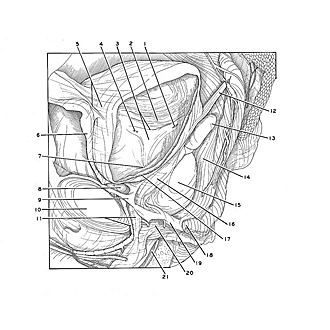
Dissection of male pelvis from a lateral approach
Interior of bladder
Image #170-6
KEYWORDS: Urinary tract.
Creative Commons
Stanford holds the copyright to the David L. Bassett anatomical images and has assigned Creative Commons license Attribution-Share Alike 4.0 International to all of the images.

For additional information regarding use and permissions, please contact the Medical History Center.

Dissection of male pelvis from a lateral approach
Interior of bladder
A large opening has been made in the left wall of the bladder to afford a view of the interior, particularly the area of the trigone.
- Left ureteric opening
- Fundus of bladder (mucosal tunic intact)
- Right pointer: Interureteric fold Left pointer: Trigone of urinary bladder (pointer indicates central part of trigone)
- Right utereric opening
- Right pointer: Apex of bladder Left pointer: Umbilicovesical fascia
- Umbilical prevesical fascia
- Internal urethral opening
- Retropubic space
- Puboprostatic ligament
- Pubic symphysis
- Upper pointer: Transverse perineal ligament Lower pointer: Dorsal vein of the penis (passing inward to prostatic plexus)
- Ureter left (cut off)
- Seminal vesicle
- Rectum
- Prostate (pointer on base)
- Pubovesicalis muscle
- Muscular tunic of bladder
- Levator ani muscle (medial border of puborectalis muscle)
- Superior fascia of urogenital diaphragm
- Sphincter muscle of urethra (remnant surrounding wall of membranous part of urethra) 21. Spongy part of urethra (opened)