Bassett Collection of Stereoscopic Images of Human Anatomy
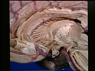
Exploration of the brain from the medial aspect
Internal capsule lateral to caudate nucleus; superior occipitofrontal fasciculus; substantia nigra
Image #17-1
KEYWORDS: Brain, Diencephalon, Midbrain, Telencephalon.
Creative Commons
Stanford holds the copyright to the David L. Bassett anatomical images and has assigned Creative Commons license Attribution-Share Alike 4.0 International to all of the images.

For additional information regarding use and permissions, please contact the Medical History Center.

Exploration of the brain from the medial aspect
Internal capsule lateral to caudate nucleus; superior occipitofrontal fasciculus; substantia nigra
The head and body of the caudate nucleus have been scraped away to demonstrate discret bundles of fibers in the internal capsule. The majority of these medially located fibers appear to extend upward from the thalamic region and turn anteriorly to form the superior occipitofrontal fasciculus located just beneath the corpus callosum. This commonly used name for this association bundle is apparently a misnomer inasmuch as none of the fibers appear related to structures in the occipital region.
- Cingulum
- Corpus callosum (cut back)
- Superior occipitofrontal fasciculus
- Stria terminalis
- Internal capsule
- Broken ends of fibers passing from head of caudate nucleus into internal capsule
- Anterior commissure
- Fibers of rostrum corpus callosum extending into frontal lobe
- Anterior cerebral artery (cut off)
- Upper pointer: Area formerly occupied by lateral hypothalamic structures Lower pointer: Optic chiasm
- Corpus callosum (splenium)
- Choroid plexus lateral ventricle and crus of fornix (cut across)
- Ends of fibers forming thalamic radiation
- Internal capsule
- Pulvinar (surface)
- Centrum medianum on cut surface of thalamus
- Superior colliculus (cut in a parasagittal plane)
- Ansa lenticularis
- Substantia nigra
- Medial lemniscus (cut across)
- Cerebral peduncle
- Oculomotor nerve (III)
- Posterior cerebral artery right