Bassett Collection of Stereoscopic Images of Human Anatomy
Dissection of male pelvis from a lateral approach
Pelvic diaphragm, close-up view of left side
Image #169-1
KEYWORDS: Muscles and tendons.
Creative Commons
Stanford holds the copyright to the David L. Bassett anatomical images and has assigned Creative Commons license Attribution-Share Alike 4.0 International to all of the images.

For additional information regarding use and permissions, please contact the Medical History Center.

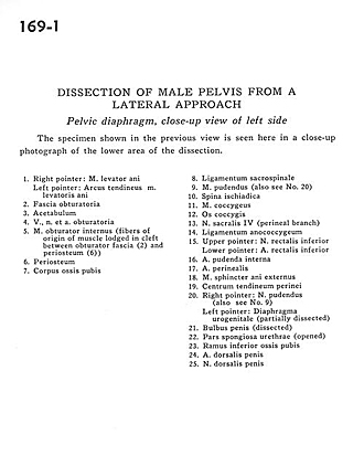
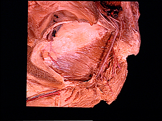
Dissection of male pelvis from a lateral approach
Pelvic diaphragm, close-up view of left side
The specimen shown in the previous view is seen here in a close-up photograph of the lower area of the dissection.
- Right pointer: Levator ani muscle Left pointer: Tendinous arch of levator ani muscle
- Obturator fascia
- Acetabulum
- Obturator nerve, artery, and vein
- Obturator internus muscle (fibers of origin of muscle lodged in cleft between obturator fascia (2) and periosteum (6)
- Periosteum
- Body of pubic bone
- Sacrospinous ligament
- Pudendal nerve (also see no. 20)
- Ischial spine
- Coccygeus muscle
- Coccyx
- Sacral nerve IV (perineal branch)
- Anococcygeal ligament
- Upper pointer: Inferior rectal nerve Lower pointer: Inferior rectal artery
- Internal pudendal artery
- Perineal artery
- External anal sphincter muscle
- Central tendon of perineum
- Right pointer: Pudendal nerve (also see no. 9) Left pointer: Urogenital diaphragm (partially dissected)
- Bulb of penis (dissected)
- Spongy part of urethra (opened)
- Inferior pubic ramus
- Dorsal artery of penis
- Dorsal nerve of the penis