
Bassett Collection of Stereoscopic Images of Human Anatomy
Male external genitalia and perineum
Contents of superficial perineal space
Image #166-5
KEYWORDS: Muscles and tendons.
Creative Commons
Stanford holds the copyright to the David L. Bassett anatomical images and has assigned Creative Commons license Attribution-Share Alike 4.0 International to all of the images.

For additional information regarding use and permissions, please contact the Medical History Center.

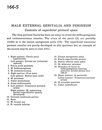
Male external genitalia and perineum
Contents of superficial perineal space
The deep perineal fascia has been cut away to reveal the bulbospongiosus and ischiocavernosus muscles. The crura of the penis (5) are partially visible as is the corpus spongiosum penis (14). The superficial transverse perineal muscles are poorly developed in this specimen but an example of the muscle may be be seen in view 174-1.
- Right pointer: Superficial penile fascia Left pointer: Scrotal sac (retracted upward)
- Deep (Buck's) fascia of penis
- Bulbospongiosus muscle
- Ischiocavernosus muscle
- Right pointer: Crus of penis Left pointer: Ramus of ischium
- Pectineus muscle
- Ischial tuberosity
- Ischiorectal fossa
- Left pointer: Transverse perineal artery Right pointer: Superficial transverse perineal muscle (poorly developed)
- Central tendon of perineum
- Anus
- Levator ani muscle
- Inferior rectal nerve
- Corpus spongiosum of penis
- Superficial perineal fascia
- Inferior pubic ramus
- Obturator externus muscle
- Branches of nerve to bulbospongiosus entering posterior scrotal plexus (see 166-3)
- Upper pointer: Perineal artery Lower pointer: Transverse perineal artery
- Posterior scrotal nerve
- Ischial tuberosity