Bassett Collection of Stereoscopic Images of Human Anatomy
Male external genitalia and perineum
Superficial fascia reflected; posterior scrotal nerves and vessels; deep perineal fascia
Image #166-3
KEYWORDS: Peripheral nervous system, Vasculature.
Creative Commons
Stanford holds the copyright to the David L. Bassett anatomical images and has assigned Creative Commons license Attribution-Share Alike 4.0 International to all of the images.

For additional information regarding use and permissions, please contact the Medical History Center.


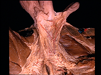
Male external genitalia and perineum
Superficial fascia reflected; posterior scrotal nerves and vessels; deep perineal fascia
The dartos has been split close to the midline at the base of the scrotum. The incision has been extended posteriorly through the membranous layer of superficial fascia (Colles' fascia). This layer has been retracted on the left side to reveal the major branches of the posterior scrotal nerves and artery. The perineal branch of the posterior femoral cutaneous nerve (10) passes anteriorly to join the plexiform branches of the posterior scrotal nerves (13). Note also that the posterior scrotal nerves communicate across the midline with branches derived from nerves of the opposite side. The adductor muscles of the left thigh have been cut away to expose the obturator externus (15). The rami of the pubis and ischium have been stripped of periosteum on the left.
- Body of penis (covered by superficial penile fascia)
- Dartos fascia (enclosing right testis)
- Septum of scrotum (compressed against penis and divided in reflecting parts of scrotum)
- Deep perineal fascia (Buck's fascia)
- Superficial perineal fascia (reflected laterally)
- Gracilis muscle
- Posterior border of area enclosed by superficial perineal fascia
- Anus
- Perineal branch of inferior rectal nerve
- Perineal branch posterior femoral cutaneous nerve
- Ischial tuberosity
- Dartos fascia (enclosing atrophic left testis)
- Branches of posterior scrotal nerves
- Superficial perineal fascia
- Obturator externus muscle
- Posterior scrotal branch internal pudendal artery
- Ramus of ischium (periosteum removed)