
Bassett Collection of Stereoscopic Images of Human Anatomy
Dissection of female pelvis from a lateral approach
Interior of left side of pelvic cavity.
Image #162-5
KEYWORDS: Ovary, Muscles and tendons, Uterus.
Creative Commons
Stanford holds the copyright to the David L. Bassett anatomical images and has assigned Creative Commons license Attribution-Share Alike 4.0 International to all of the images.

For additional information regarding use and permissions, please contact the Medical History Center.

Dissection of female pelvis from a lateral approach
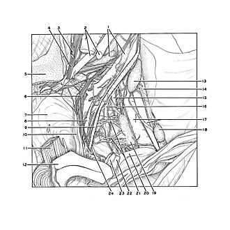
Interior of left side of pelvic cavity.
Removal of the left ovary and uterine tube has exposed the structures that lie immediately lateral to the ovarian fossa. Lymphatic vessels (21) can be seen passing to the external iliac nodes from the uterus and vagina. The obliterated part of the umbilical artery (16) lies lateral to these vessels against the pelvic wall.
- Iliac lymph nodes
- Right pointer: External iliac artery (cut off) Left pointer: Internal iliac artery
- Femoral nerve
- Obturator nerve
- Articular surface of sacrum
- Superior hypogastric plexus
- Parietal pelvic fascia
- Uterine vein
- Ureter
- Rectouterine muscle (uterosacral ligament)
- Rectum (cut across)
- Uterus
- External iliac lymph node
- Psoas major muscle (covered by fascia)
- Obturator artery
- Lateral umbilical ligament
- External iliac artery and vein
- External iliac lymph node
- Internal iliac lymph node
- Ligamentum teres (of uterus)
- Lymph vessel (from uterus and vagina)
- Uterine tube (cut across)
- Proper ovarian ligament (cut off)
- Uterine artery