
Bassett Collection of Stereoscopic Images of Human Anatomy
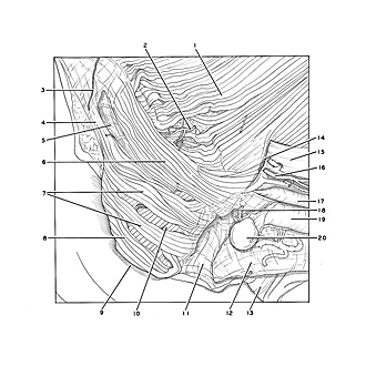
Dissection of female pelvis froma lateral approach
Sphincter ani externus muscle, close-up view
Image #159-6
KEYWORDS: Muscles and tendons, Bones joints cartilage.
Creative Commons
Stanford holds the copyright to the David L. Bassett anatomical images and has assigned Creative Commons license Attribution-Share Alike 4.0 International to all of the images.

For additional information regarding use and permissions, please contact the Medical History Center.

Dissection of female pelvis froma lateral approach
Sphincter ani externus muscle, close-up view
The specimen shown in the previous view is here seen in a close-up centered on the sphincter ani externus muscle.
- Levator ani muscle (pointer on puborectalis muscle)
- Inferior rectal artery (small branch cut off)
- Anococcygeal nerve
- Anococcygeal ligament
- Area of communication between ischiorectal fossae of two sides
- Deep part
- Superficial part
- Subcutaneous part (6-8 refer to the external anal sphincter)
- Anus
- Longitudinal muscle layer of anal wall
- Central tendon of perineum
- Vaginal wall
- Vestibule of vagina (opened by removal of labia on right side)
- Ischiorectal fossa (remnant of anterior recess)
- Obturator fascia
- Dorsal artery of clitoris
- Crus of clitoris
- artery of vestibular bulb
- Vestibular bulb
- Major vestibular gland