
Bassett Collection of Stereoscopic Images of Human Anatomy
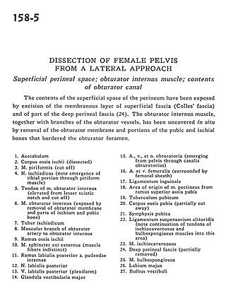
Dissection of female pelvis from a lateral approach
Superficial perineal space; obturator internus muscle; contents of obturator canal
Image #158-5
KEYWORDS: Muscles and tendons, Perineum, Bones joints cartilage, Urinary tract.
Creative Commons
Stanford holds the copyright to the David L. Bassett anatomical images and has assigned Creative Commons license Attribution-Share Alike 4.0 International to all of the images.

For additional information regarding use and permissions, please contact the Medical History Center.

Dissection of female pelvis from a lateral approach
Superficial perineal space; obturator internus muscle; contents of obturator canal
The contents of the superficial space of the perineum have been exposed by excision of the membranous layer of superficial fascia (Colles' fascia) and of part of the deep perineal fascia (24). The obturator internus muscle, together with branches of the obturator vessels, has been uncovered in situ by removal of the obturator membrane and portions of the pubic and ischial bones that bordered the obturator foramen.
- Acetabulum
- Body of ischium (dissected)
- Piriform muscle (cut off)
- Sciatic nerve (note emergence of tibial portion through piriform muscle)
- Tendon of obturator internus muscle (elevated from lesser sciatic notch and cut off)
- Obturator internus muscle (exposed by removal of obturator membrane and parts of ischium and pubic bone)
- Ischial tuberosity
- Muscular branch of obturator artery to obturator internus
- Ramus of ischium
- External anal sphincter muscle (muscle fibers indistinct)
- Posterior labial branch internal pudendal artery
- Posterior labial nerve
- Posterior labial vein (plexiform)
- Major vestibular gland
- Obturator nerve, artery, and vein (emerging from pelvis through obturator canal)
- Femoral artery and vein (surrounded by femoral sheath)
- Inguinal ligament
- Area of origin of pectineus muscle from superior pubic ramus
- Pubic tubercle
- Body of pubic bone (partially cut away)
- Pubic symphysis
- suspensory ligament of clitoris (note continuation of tendons of ischiocavernosus and bulbospongiosus muscles into this area)
- Ischiocavernosus muscle
- Deep perineal fascia (partially removed)
- bulbospongiosus muscle
- Labium majus
- Vestibular bulb