
Bassett Collection of Stereoscopic Images of Human Anatomy
Dissection of female pelvis from a lateral approach
Right obturator internus muscle, general lateral view
Image #158-4
KEYWORDS: Muscles and tendons, Bones joints cartilage.
Creative Commons
Stanford holds the copyright to the David L. Bassett anatomical images and has assigned Creative Commons license Attribution-Share Alike 4.0 International to all of the images.

For additional information regarding use and permissions, please contact the Medical History Center.

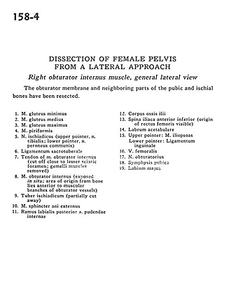
Dissection of female pelvis from a lateral approach
Right obturator internus muscle, general lateral view
The obturator membrane and neighboring parts of the pubic and ischial bones have been resected.
- Gluteus minimus muscle
- Gluteus medius muscle
- Gluteus maximus muscle
- Piriform muscle
- Sciatic nerve (upper pointer, tibial nerve; lower pointer, common peroneal nerve)
- Sacrotuberous ligament
- Tendon of obturator internus muscle (cut off close to lesser sciatic foramen gemelli muscles removed)
- Obturator internus muscle (exposed in situ area of origin from bone lies anterior to muscular branches of obturator vessels)
- Ischial tuberosity (partially cut away)
- External anal sphincter muscle
- Posterior labial branch internal pudendal artery
- Body of ilium
- Anterior inferior iliac spine (origin of rectus femoris visible)
- Acetabular labrum
- Upper pointer: Iliopsoas muscle Lower pointer: Inguinal ligament
- Femoral vein
- Obturator nerve
- Pubic symphysis
- Labium majus