Bassett Collection of Stereoscopic Images of Human Anatomy
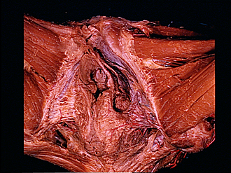
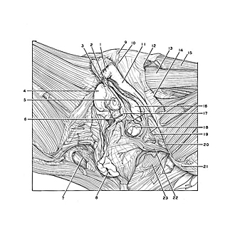
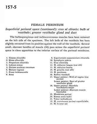
Female perineum
Superficial perineal space (continued).
Image #157-5
KEYWORDS: Muscles and tendons, Perineum, Bones joints cartilage, Urinary tract.
Creative Commons
Stanford holds the copyright to the David L. Bassett anatomical images and has assigned Creative Commons license Attribution-Share Alike 4.0 International to all of the images.

For additional information regarding use and permissions, please contact the Medical History Center.
Female perineum
Superficial perineal space (continued).
The bulbospongiosus and ischiocavernosus muscles have been removed on the left side of the specimen. The left bulb of the vestibule has been slightly retracted from its position against the wall of the vestibule. Several small, aberrant bundles of muscle (18) pass across the superficial perineal space in close apposition to the inferior surface of the perineal membrane.
- Body of clitoris
- Glans of clitoris
- Prepuce of clitoris
- Labium minus
- External urethral opening
- Vaginal opening
- Ischiorectal fossa
- Anus
- Suspensory ligament of clitoris
- Pubic symphysis
- Crus of clitoris
- Adductor longus muscle (cut off)
- Adductor brevis muscle
- Gracilis muscle (cut off)
- Pectineus muscle
- Bulbus vestibuli
- Upper pointer: Wall of vagina (cut across) Lower pointer: Duct of greater vestibular gland
- Upper pointer: Greater vestibular gland Lower pointer: Aberrant muscle fasciculus
- Perineal membrane
- Ramus of ischium
- Ischial tuberosity
- Superficial transverse perineal muscle
- Levator ani muscle