Bassett Collection of Stereoscopic Images of Human Anatomy
Female perineum
Superficial perineal space (continued).
Image #157-4
KEYWORDS: Muscles and tendons, Perineum, Peripheral nervous system, Bones joints cartilage, Central nervous system, Urinary tract.
Creative Commons
Stanford holds the copyright to the David L. Bassett anatomical images and has assigned Creative Commons license Attribution-Share Alike 4.0 International to all of the images.

For additional information regarding use and permissions, please contact the Medical History Center.
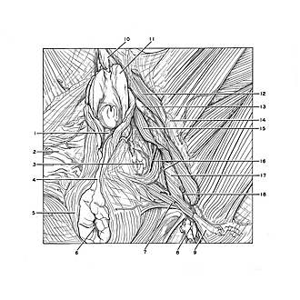
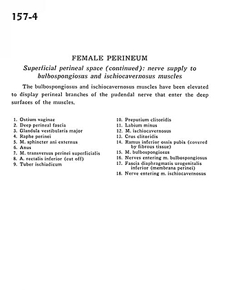
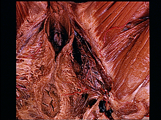
Female perineum
Superficial perineal space (continued).
The bulbospongiosus and ischiocavernosus muscles have been elevated to display perineal branches of the pudendal nerve that enter the deep surfaces of the muscles.
- Vaginal opening
- Deep perineal fascia
- Greater vestibular (Bartholin's) gland
- Perineal raphe
- External anal sphincter muscle
- Anus
- Superficial transverse perineal muscle
- Inferior rectal artery (cut off)
- Ischial tuberosity
- prepuce of clitoris
- Labium minus
- Ischiocavernosus muscle
- Crus of clitoris
- Inferior pubic ramus (covered by fibrous tissue)
- Bulbospongiosus muscle
- Nerves entering bulbospongiosus muscle
- Inferior fascia of urogenital diaphragm (perineal membrane)
- Nerve entering ischiocavernosus muscle