Bassett Collection of Stereoscopic Images of Human Anatomy
Ligaments and joints of lumbosacral spine and pelvic girdle
Sagittal section of lumbosacral spine; transverse section through sacroiliac joint
Image #156-1
KEYWORDS: Central nervous system, Bones joints cartilage, Vasculature.
Creative Commons
Stanford holds the copyright to the David L. Bassett anatomical images and has assigned Creative Commons license Attribution-Share Alike 4.0 International to all of the images.

For additional information regarding use and permissions, please contact the Medical History Center.
Ligaments and joints of lumbosacral spine and pelvic girdle
Sagittal section of lumbosacral spine; transverse section through sacroiliac joint
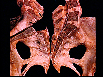
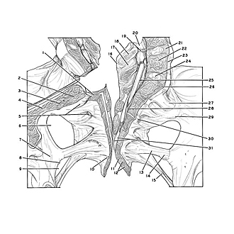
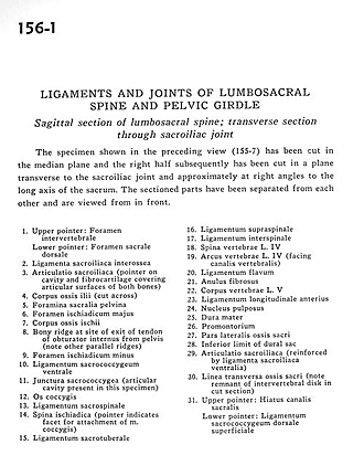
The specimen shown in the preceding view (155-7) has been cut in the median plane and the right half subsequently has been cut in a plane transverse to the sacroiliac joint and approximately at right angles to the long axis of the sacrum. The sectioned parts have been separated from each other and are viewed from in front.
- Upper pointer: Intervertebral foramen Lower pointer: Dorsal sacral foramen
- Interosseous sacroiliac ligament
- Sacroiliac joint (pointer on cavity and fibrocartilage covering articular surfaces of both bones)
- Body of ilium (cut across)
- Anterior (pelvic) sacral foramina
- Greater sciatic foramen
- Body of ischium
- Bony ridge at site of exit of tendon of obturator internus from pelvis (note other parallel ridges)
- Lesser sciatic foramen
- Ventral sacrococcygeal ligament
- Sacrococcygeal junction (articular cavity present in this specimen)
- Coccyx
- Sacrospinous ligament
- Ischial spine (pointer indicates facet for attachment of coccygeus muscle)
- Sacrotuberous ligament
- Supraspinous ligament
- Interspinous ligament
- Spine of vertebra L. IV
- Arch of vertebra L. IV (facing vertebral canal)
- Ligamentum flavum
- Anulus fibrosus
- Body of vertebra L. V
- Anterior longitudinal ligament
- Nucleus pulposus
- Dura mater
- Promontory
- Lateral part of sacrum
- Inferior limit of dural sac
- Sacroiliac joint (reinforced by ventral sacroiliac ligament)
- Transverse line of sacrum (note remnant of intervertebral disk in cut section)
- Upper pointer: Hiatus sacral canal Lower pointer: Superficial dorsal sacrococcygeal ligament