Bassett Collection of Stereoscopic Images of Human Anatomy
Osteology
Articulated male and female pelvic bones, anterior view
Image #153-1
KEYWORDS: Bones joints cartilage, Muscles and tendons, Vasculature.
Creative Commons
Stanford holds the copyright to the David L. Bassett anatomical images and has assigned Creative Commons license Attribution-Share Alike 4.0 International to all of the images.

For additional information regarding use and permissions, please contact the Medical History Center.

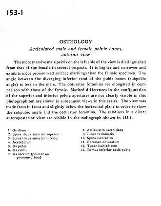
Osteology
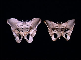
Articulated male and female pelvic bones, anterior view
The more massive male pelvis on the left side of the view is distinguished from that of the female in several respects. It is higher and narrower and exhibits more pronounced surface markings than the female specimen. The angle between the diverging inferior rami of the pubic bones (subpubic angle) is less in the male. The obturator foramina are elongated in comparison with those of the female. Marked differences in the configuration of the superior and inferior pelvic apertures are not clearly visible in this photograph but are shown in subsequent views in this series. The view was made from in front and slightly below the horizontal plane in order to show the subpubic angle and the obturator foramina. The relations in a direct anteroposterior view are visible in the radiograph shown in 155-1.
- Ilium
- Anterior superior iliac spine
- Anterior inferior iliac spine
- Acetabulum
- Pubic bone
- Ischium
- Sacrum (pointer on Promontory)
- Sacroiliac joint
- Linea terminalis
- Ischial spine
- Obturator foramen
- Ischial tuberosity
- Inferior pubic ramus