
Bassett Collection of Stereoscopic Images of Human Anatomy
Kidneys, suprarenal glands and posterior abdominal vessels, nerves and muscles
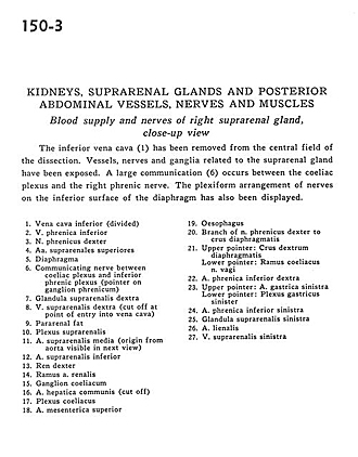
Blood supply and nerves of right suprarenal gland, close-up view
Image #150-3
KEYWORDS: Adrenal gland, Kidney, Muscles and tendons, Peripheral nervous system, Vasculature.
Creative Commons
Stanford holds the copyright to the David L. Bassett anatomical images and has assigned Creative Commons license Attribution-Share Alike 4.0 International to all of the images.

For additional information regarding use and permissions, please contact the Medical History Center.

Kidneys, suprarenal glands and posterior abdominal vessels, nerves and muscles
Blood supply and nerves of right suprarenal gland, close-up view
The inferior vena cava (1) has been removed from the central field of the dissection. Vessels, nerves and ganglia related to the suprarenal glands have been exposed. A large communication (6) occurs between the coeliac plexus and the right phrenic nerve. The plexiform arrangement of nerves on the inferior surface of the diaphragm has also been displayed.
- Inferior vena cava (divided)
- Inferior phrenic vein
- Right phrenic nerve
- Superior suprarenal arteries
- Diaphragm
- Communicating nerve between celiac plexus and inferior phrenic plexus (pointer on phrenic ganglion)
- Right suprarenal gland
- Right suprarenal vein (cut off at point of entry into vena cava)
- Pararenal fat
- Suprarenal plexus
- Middle suprarenal artery (origin from aorta visible in next view)
- Inferior suprarenal artery
- Right kidney
- Branch of renal artery
- Celiac ganglion
- Common hepatic artery (cut off)
- Celiac plexus
- Superior mesenteric artery
- Esophagus
- Branch of right phrenic nerve to crus of diaphragm
- Upper pointer: Right crus of diaphragm Lower pointer: Celiac branch of vagus nerve
- Right inferior phrenic artery
- Upper pointer: Left gastric artery Lower pointer: Left gastric plexus
- Left inferior phrenic artery
- Left suprarenal gland
- Splenic artery
- Left suprarenal vein