Bassett Collection of Stereoscopic Images of Human Anatomy
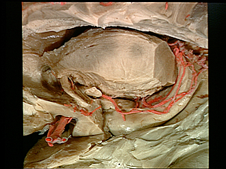
Exploration of the brain from its lateral aspect
Relations of inferior horn of lateral ventricle; choroidal arteries
Image #15-3
KEYWORDS: Brain, Diencephalon, Telencephalon, Vasculature, Ventricules.
Creative Commons
Stanford holds the copyright to the David L. Bassett anatomical images and has assigned Creative Commons license Attribution-Share Alike 4.0 International to all of the images.

For additional information regarding use and permissions, please contact the Medical History Center.

Exploration of the brain from its lateral aspect
Relations of inferior horn of lateral ventricle; choroidal arteries
The lateral geniculate body, sublenticular part of the internal capsule and the optic tract have been cut away to reveal the course of the choroidal vessels. The choroid plexus has been dissected away in order to expose the vessels. The fornix has been exposed further by cutting away parts of the pulvinar and posterior thalamic radiation.
- Septum pellucidum
- Stria terminalis
- Internal capsule (cut across)
- Radiation of rostral lamina of corpus callosum
- Area of internal capsule related to globus pallidus (the latter has been removed)
- Anterior commissure
- Optic tract (cut across)
- Choroidal artery (anterior)
- Limen insulae (cut across lateral to substantia perforata anterior)
- Amygdaloid nucleus
- Middle cerebral artery
- Corpus callosum (cut across)
- External medullary lamina (thalamus) (cut across)
- Pulvinar (cut across)
- Fimbria of hippocampus
- Cerebral peduncle
- Hippocampal gyrus
- Uncus (hippocampal gyrus)
- Hippocampus
- Hippocampal digitations
- Medullary substance of temporal lobe