Bassett Collection of Stereoscopic Images of Human Anatomy
Exploration of liver, gall bladder, pancreas, duodenum and spleen
Interior of gall bladder, bile ducts, close-up view
Image #146-7
KEYWORDS: Gallbladder, Liver, Pancreas, Spleen.
Creative Commons
Stanford holds the copyright to the David L. Bassett anatomical images and has assigned Creative Commons license Attribution-Share Alike 4.0 International to all of the images.

For additional information regarding use and permissions, please contact the Medical History Center.
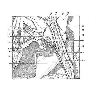
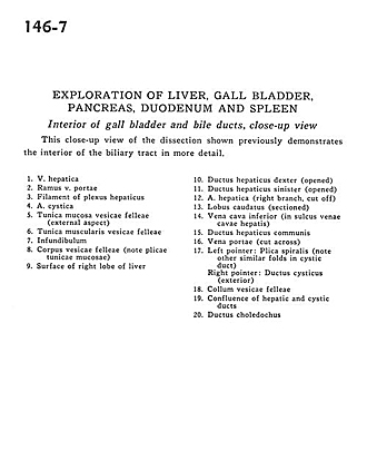
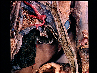
Exploration of liver, gall bladder, pancreas, duodenum and spleen
Interior of gall bladder, bile ducts, close-up view
This close-up view of the dissection shown previously demonstrates the interior of the biliary tract in more detail.
- Hepatic vein
- Branch of portal vein
- Filament of hepatic plexus
- Cystic artery
- Muscular layer of gallbladder (external aspect)
- Muscular layer of gallbladder
- Infundibulum
- Body of gallbladder (note plicae tunicae mucosae)
- Surface of right lobe of liver
- Right hepatic duct (opened)
- Left hepatic duct (opened)
- Hepatic artery (right branch, cut off)
- Caudate lobe (sectioned)
- Inferior vena cava (in sulcus of vena cava hepatis)
- Common hepatic duct
- Portal vein (cut across)
- Left pointer: Spiral folds (note other similar folds in cystic duct) Right pointer: Cystic duct (exterior)
- Neck of gallbladder
- Confluence of hepatic and cystic ducts
- Common bile duct