Bassett Collection of Stereoscopic Images of Human Anatomy
Exploration of liver, gall bladder, pancreas, duodenum and spleen
Dissection of pancreas (continued).
Image #146-4
KEYWORDS: Gallbladder, Liver, Pancreas, Spleen.
Creative Commons
Stanford holds the copyright to the David L. Bassett anatomical images and has assigned Creative Commons license Attribution-Share Alike 4.0 International to all of the images.

For additional information regarding use and permissions, please contact the Medical History Center.
Exploration of liver, gall bladder, pancreas, duodenum and spleen
Dissection of pancreas (continued).
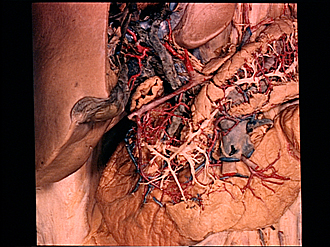
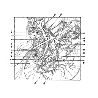
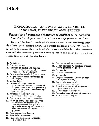
Some of the blood vessels which were shown in the preceding dissection have been cleared away. The gastroduodenal artery (5) has been retracted to expose the area in which the common bile duct, the pancreatic duct and the accessory pancreatic duct approach and enter the wall of the descending part of the duodenum.
- Cystic artery
- Cystic duct
- Junction of cystic and hepatic ducts to form common bile duct (traceable to duodenum)
- Superior part of duodenum (cut across)
- Gastroduodenal artery (retracted to the right)
- Gallbladder
- Lowest of three superior pancreaticoduodenal branches of gastroduodenal artery (in preceding view this branch is indicated by pointer #22)
- Arterial arches between pancreaticoduodenal arteries
- Accessory pancreatic duct
- Point of entry into duodenal wall of common bile duct and pancreatic duct (in this specimen these open separately into duodenum at duodenal papilla)
- Descending part of duodenum
- Inferior part of duodenum
- Uncinate process of pancreas (partially dissected)
- Common hepatic duct
- Upper pointer: Proper hepatic artery Lower pointer: Portal vein
- Splenic artery
- Pancreatic duct
- Splenic vein
- Body of pancreas (intact portion of upper border)
- Pancreatic branch of superior mesenteric artery
- Junction of main and accessory pancreatic ducts
- Superior mesenteric vein
- Posterior part of head of pancreas
- Superior mesenteric artery