Bassett Collection of Stereoscopic Images of Human Anatomy
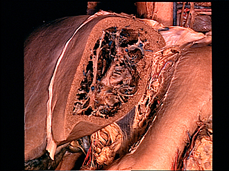
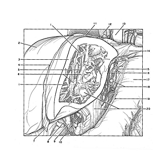
Exploration of liver, gall bladder, pancreas, duodenum and spleen
Dissection of left lobe of liver, viewed from left
Image #145-1
KEYWORDS: Gallbladder, Liver, Pancreas, Spleen.
Creative Commons
Stanford holds the copyright to the David L. Bassett anatomical images and has assigned Creative Commons license Attribution-Share Alike 4.0 International to all of the images.

For additional information regarding use and permissions, please contact the Medical History Center.
Exploration of liver, gall bladder, pancreas, duodenum and spleen
Dissection of left lobe of liver, viewed from left
The left lobe of the liver has been cut in a saggital plane parallel to the falciform ligament. The interior of the lobe has been dissected to display branches of the hepatic artery, hepatic duct and portal vein ensheathed in a common layer of fibrous tissue (6). These structures comprise the portal triad and occupy an interolobular position. Tributaries of one of the hepatic veins (1), centrally located with respect to the lobules, also may be traced in the dissection. These converge toward the vena cava and are better seen in later stages of the dissection. The extension of the lesser omentum (20) into the depths of the fissure of the ligamentum venosum is illustrated.
- Hepatic vein
- Right lobe of liver
- Left lobe of liver (sectioned in sagittal plane)
- Falciform ligament
- Ligamentum venosum (in depth of dissected area)
- Perivascular fibrous capsule (surrounding branches of portal vein, hepatic artery and bile duct; upper pointer, superior lateral segmental branches of left lobe; lower pointer, inferior lateral segmental branches of (left lobe)
- Ligamentum teres
- Peritoneum at margin of dissected lesser omentum
- Common bile duct
- Hepatic lymph node
- Coronary ligament (diaphragm divided close to attachment of ligament)
- Esophagus
- Thoracic aorta
- Diaphragm
- Abdominal part of esophagus
- Filament of gastric plexus
- Stomach
- Left gastric lymph nodes
- Peritoneum cut at margin of dissected lesser omentum
- Lesser omentum (upper pointer indicates part within depths of fissure of ligamentum venosum; lower pointer indicates undissected part of lesser omentum)